Parkinson’s Beyond the Brain

Photo credit to Wikimedia Commons

By Emma Yasinski
BU News Service

By the time a patient is diagnosed with Parkinson’s disease, seventy percent of the damage is already done. In an effort to understand the progression of the disease and diagnose it earlier, more and more scientists are moving their microscopes to the patient’s gut.

Researchers agree that the most characteristic traits of Parkinson’s disease, difficulties with movement, are caused by protein clumps found in the substantia nigra, a small area deep in the brain. In 2003, renowned researcher, Heiko Braak, PhD at Johann Wolfgang Goethe University in Germany, proposed a theory that rather than starting in the brain, the disease might start elsewhere in the body. Based on patterns he observed in autopsies, he proposed that alpha-synuclein, the protein considered to be the main cause of damage in the Parkinson’s brain, might spread from nerve cell to nerve cell. He suggested the protein might start outside the brain and travel through nerve cells up to the substantia nigra. Before this, most researchers believed that these proteins aggregated only in the substantia nigra- and nowhere else. His proposal completely changed the way researchers thought about the progression of the disease.

Dr. Kathleen Shannon of Rush University Medical Center in Chicago was in the middle of reading Braak’s 2003 study when she received an email from her new collaborator, a gastroenterologist. He was at a conference in another state, learning about Parkinson’s because a family member had been recently diagnosed with the disease. He perked up when he heard researchers describe a new rodent model for the disease. The model used a neurotoxin called lipopolysaccharide to induce symptoms. As a gastroenterologist, he recognized the toxin. “Did you know the only route of exposure is through the intestinal system?” His email to Shannon stated that this was their next clue to a new understanding of the disease. If this toxin is entering through the intestines and leading to Parkinson’s-like symptoms, maybe the real disease pathology works this way too.

They immediately recruited nine patients who were recently diagnosed with Parkinson’s, but had not yet begun treatment. The first goal was to test whether the patients had a “leaky gut” – a condition in which pathogens are able to move across the intestinal wall more easily than usual. They gave the patients several different types of sugars and then tested for their presence in the patients’ urine. Depending on the ratio of different sized sugars found in the urine, the researchers were able to tell how permeable, or “leaky” each patient’s colon was. Those that had been recently diagnosed with Parkinson’s showed evidence of significantly leakier guts than healthy patients.The patients’ intestines also showed evidence of alpha-synuclein, the protein most closely associated with Parkinson’s disease. The researchers hypothesized that the body might start producing protein in response to a toxin that enters through the leaky intestinal walls.

The following year the researchers tracked down biopsies from three of their patients’ past colonoscopies. The researchers’ goal was to see if alpha-synuclein was present in the intestines prior to the onset of Parkinson’s symptoms. The samples came from 2-5 years before the individuals were diagnosed with Parkinson’s. All three patients’ biopsies showed alpha-synuclein in the colon. In contrast, she found no evidence of alpha-synuclein in the colons of the 23 healthy patients. This was the first study showing that the protein could be found in the gut prior to a patient’s diagnosis.

Several teams of researchers are currently working to validate this finding with larger sample sizes. If they find the same results, doctors could diagnose Parkinson’s disease earlier than ever, during a routine colonoscopy. Earlier diagnosis of the disease would make it possible to begin treatment earlier before most of the neurons are already damaged. Right now, doctors can only diagnose the disease when the patient displays motor symptoms. By then, 70% of the neurons in the substantia nigra are dead. It’s possible that an ability to diagnose the disease earlier would allow researchers to intervene, and maybe even find a way to slow or prevent the cell death.

Early diagnosis is only one benefit to elucidating the roots of alpha-synuclein in the body. As researchers gain a better understanding of Parkinson’s, Shannon hopes they can work backwards to create better animal models. She explained that current animal models only mimic the disease’s effects on the brain, and not the rest of the body. Researchers create one of the most common models by damaging the neurons in the substantia nigra of rats’ brains. If scientists hope to develop treatments that target pathology or symptoms outside of the brain, they will need more accurate models, which mimic the disease pathology throughout the body. Beth Vernaleo, PhD., Senior Manager of Research Programs at the Parkinson’s Disease Foundation, described a lack of accurate animal models as one of the greatest challenges facing Parkinson’s researchers. “You have to start there.” She said “When you’re looking at things like colonoscopy samples, you’re hypothesizing what’s happened. You have to look back at biological models to see if it’s actually what is happening.”

Some researchers still aren’t convinced. Thomas Beach, PhD, Director of the Brain and Body Donation Program at Banner Sun Health Research Institute, does not believe that Parkinson’s disease pathology starts outside of the brain at all. His research, based on an extensive collection of autopsies, suggests that while the patient may not have symptoms until the pathology has reached other areas of the body, it still begins in the brain, “We have never detected alpha-synuclein deposits in the body unless they were already somewhere in the brain,” he said “It does not start in the body. It starts in the brain and moves to the body after.” He does, however, agree that the alpha synuclein deposits throughout the body could be useful for confirming diagnosis and treating specific symptoms of the illness.

Whether scientists hope to find a diagnostic marker, a treatment, or an animal model, it seems most researchers and patients agree that looking to the intestines is an important new step in Parkinson’s disease research. And the trend is catching on. Researchers specializing in other neurological diseases, such as Multiple Sclerosis and Alzheimer’s disease, are following suit and looking at changes in the gut to understand the illnesses. A group of researchers at several different institutions across the United States has come together to create the Multiple Sclerosis Microbiome Consortium (MSMC) to share information on the topic. Charlotte Madore, PhD at Brigham and Women’s Hospital, has just started a study about the relationship between the gut microbiota and cells that lead to inflammation in the brain. If there is a relationship between the two, it will be a crucial piece of the puzzle not just in Parkinson’s, but all neurodegenerative diseases.

As of right now, doctors have no way to stop the progression of these diseases; all they can do is temporarily alleviate the symptoms. Perhaps the keys to reversing them lie in the gut.

Decreasing Deadly Desensitization

Image by Kalumet.
Image by Kalumet.

By Emma Yasinski
BU News Service

Walking past patient’s rooms on the cardiac floor of Boston Medical Center, I could hear nurses ruffling papers at the central station. The wheels of a monitor scratched against the hard, white floors. Nurses and doctors spoke at relaxed volumes. A nurse walked by and I heard a quiet beep-beep sound. “Was that an alarm?” I asked Deborah Whalen, a clinical nurse manager at Boston Medical Center. “No that’s just a beeper.” We’d been standing on the cardiac floor for 15 minutes and had yet to hear an alarm.

This hall sounded very different 15 months ago.

Nurses here used to be overwhelmed by alarms. In most other hospitals, they still are. Barbara Drew, RN, PhD, FAAN at the University of California San Francisco recently counted 2.5 million alarms during a month-long period in their Intensive Care Unit. Each patient triggered an average of 45 alarms per hour. The study also found that 88% of these alarms were false. Over time, the alarms became background noise for caretakers. “When I worked on my undergrad…Georgetown was right near a national airport. [At first] I would hear every plane landing and taking off,” said Whalen, describing her first year in a hospital setting. “Then, by the end of my freshman year, I didn’t even hear a plane. You get accustomed to the sound and therefore you block it out.” The decibel level of the background alarms was so high that not even a plane taking off would get her attention anymore.

This is the type of environment that investigators say led to the death of a patient in the cardiac unit at Massachusetts General Hospital in 2010. When there are so many alarms blaring, nurses sometimes lower their volumes, or even turn them off in an effort to reduce noise. The patient was recovering from surgery and had been hooked up to a variety of monitors. When his heart rate dropped, not one of the 10 nurses on the floor heard the alarm or came to his aid. The investigation showed that the alarm didn’t sound at all. A nurse had purposely turned the alarm off.

The Joint Commission, a group responsible for accrediting hospitals, listed alarm fatigue as a crucial problem in patient safety in 2014. There have been instances, such as that in 2010, where desensitization to alarms led to disastrous consequences.The Joint Commission reported 80 deaths related to alarm issues between 2009 and 2012. Reporting these events is optional for hospitals, so it is likely that the actual number of deaths related to alarms is much higher. To be accredited in 2016, hospitals will need to improve their alarm management.

Boston Medical Center had two main types of audible alarms: crisis alarms and warning alarms. The difference between the two alarms is a single beep. A warning alarm is two high-pitched beeps, while a crisis alarm is three high-pitched beeps. Each beep sounds the same. The warnings don’t usually require immediate intervention. With such a high volume of alarms, it can be difficult for nurses to distinguish between a crisis and a warning. After observing the pilot floor, Whalen and her team discovered that most of the noise was coming from warning alarms, but that nurses rarely answered them. They were much more likely to respond to the higher level crisis alarms, but the distraction and background noise from the warning alarms still distracted them and made it difficult for them to identify a true crisis.

Boston Medical Center had just begun studying the effects of alarm fatigue when the patient at Massachusetts General Hospital passed away. After determining that they too might be vulnerable to the effects of alarm fatigue, Whalen and her colleagues began developing a plan to safely reduce the noise on the hospital floors. The first change they made was removing superfluous warning alarms. The few warning alarms that were deemed urgent were upgraded to crisis alarms. Now the condition that would have triggered a two-beep warning before still showed up in the patient’s history, but do not make distracting noise. The audible alarms are reserved for true emergencies.

Next, the hospital had to decide who had the power to change the alarm settings on a specific patient and when. Every individual patient is different. A heart rate that may signal an emergency in one patient may not signal an emergency in another. For example, the hospital’s default settings will sound the alarm sound if a patient’s heart rate falls below 45 beats per minute. However, many athletes have lower-than-average heart rates when they are resting or sleeping, meaning their alarms would sound frequently, but not signify a problem. In this case, two nurses at Boston Medical Center can come together and choose to set new parameters on this individual’s monitor. When the two nurses agree on the appropriate setting, they change it and then report it to a doctor on staff. Putting this power in the hands of the nurses on staff allows for an immediate reduction in noise volume. Whalen’s reasoning for requiring two nurses to make the change is that “Two nurses together making a bad decision (knock on wood) has never happened.” These changes, along with increased training, helped reduce alarms from approximately 90,000 per week to 10,000 per week. At the end of the six week pilot, the nurses begged to keep the changes in effect.

Drew, whose study on the prevalence of alarms was funded by General Electric, is working with manufacturers to identify places where the technology can improve. “There’s a lot of things hospitals can do,” she said “but manufacturers also need to step up.” Drew argues that hospitals need smarter alarms, able to take data from several sources on a single patient, such as heart rate and oxygen levels. The alarm would be able to identify patterns that often occur together before and during emergencies. Then, only a single alarm would sound in the event of a specific emergency. However, these smarter alarms could present legal issues. Rather than reporting on specific parameters, a smart alarm is responsible for diagnosing certain conditions. This has the potential to make the alarm manufacturer liable in the event of a misdiagnosis.

Whalen, Drew, and a few others have become leaders in the field. Hospitals around the country have been requesting their help in understanding and implementing these strategies. While the Joint Commission is not requiring that alarm fatigue be reduced until 2016, it is requiring that hospitals show that they have identified it as a problem and are starting to work on it this year. “We are trying to take an incremental approach,” said Gerald Castro, MD, PhD, Project Director of Safety Initiatives at the Joint Commission. “What we want them to do is to first address the problem within their organization.” One of the greatest difficulties with addressing the problem, is that the data regarding it is incomplete. Researchers aren’t certain how often alarm fatigue leads to problems for patients, because hospitals are not required to report it. Plus, if they do choose to report it, they can send the report to any of several organizations, including the Joint Commission, the FDA, or another Patient Safety Organization. These organizations haven’t been able to combine their data yet. Castro describes this as the next big step for the Joint Commission.

As we waited on the floor, still no true alarms sounded. Whalen offered to set one off for me just so I could hear the three shrill beeps and understand what it sounded like. She did, and while several nurses looked her way, they saw her turn it off and continued about their business. We watched another nurse enter a new patient’s information into the monitor and set his parameters. She typed his last name and first initial into the small blank spaces, and clicked on options from some drop down menus. After she walked away, I heard “Beep! Beep! Beep!” Whalen turned around, scoured the monitor. She clicked a button and the alarm stopped. “That one was junk,” she said, before quietly walking me back to the elevator.

A Little Boy’s Battle for Birthday Cake

alexander pirate
Alexander, dressed as a pirate, blows out the candles on his 5th birthday cake. An intestinal transplant will help him be able to enjoy a big slice. Photo by Emma Yasinski

By Emma Yasinski
BU News Service

Four-year-old Alexander was sitting on a tricycle with one foot on each side resting against the sidewalk like Fred Flintstone when I met him. He hadn’t figured out how to put his feet on the pedals yet, but was using the bike to roll around anyway. And he wasn’t willing to share the toy with his older half-brother. Scrunching his chin against his Mickey Mouse T-shirt, he scowled as he “biked” away as fast as he could. In most ways, Alexander was just like the other boys he was playing with that day, but there was one big difference: a small plastic tube taped to his cheek leading into one nostril from a bag of liquid nutrients in his backpack.

Without this tube, Alexander would starve. It delivers Total Parenteral Nutrition (TPN,) a type of liquid nutrition which he needs to grow and survive. He doesn’t eat chicken nuggets or broccoli like most kids because he doesn’t have the intestines to digest them. Days after he was born, a severe infection attacked the tissue of his digestive system. He eventually beat the disease, but not until a surgeon removed his entire colon and 80% of his small intestine. Alexander can live with this feeding regimen for a while, but eventually it will lead to repetitive infections that send him to the hospital for days or weeks at a time. Only six veins in his body are large enough to receive the TPN. Over time, these veins can get damaged or infected and he won’t be able to receive the liquid any longer. His best chance at an almost-normal life is an intestinal transplant, and he is currently on the waiting list.

Only recently have intestinal transplants become a viable option for patients like Alexander. While other organs, such as kidneys and livers, have been successfully transplanted since the 1960s, the intestines present special difficulties. With any transplant, the patient’s body might reject the new organ, but intestinal transplants pose heightened risks. The immunosuppressant techniques that doctors use for other organ transplants have not been equally effective for intestines. The intestines contain a high concentration of immune cells and bacteria, which makes it difficult for a patient’s body to accept a replacement. Even after the transplant , Alexander will need to live near the hospital for up to a year so that doctors can monitor his immune response several times a week make sure his body is accepting the organ while protecting him against opportunistic infections. Moreover, TPN that nourishes him puts stress on his liver. The longer he waits for his transplant, the more likely it is that his liver will become damaged beyond repair. If that happens, he will need a new liver along with his new intestines.

The intestines house up to 70% of the body’s immune cells, which makes them particularly vulnerable to rejection. These cells protect the large surface area of the intestines from infections that can come from food or the outside world. Unfortunately, this same protective power can turn against the tissue of the new intestine. Since patches of immune cells are found throughout the organ, the attacks take place over its entire length. The immunosuppressant drugs that work for other types of transplants often don’t prevent rejection in intestinal transplant patients. For that reason, long after other transplants procedures had gained success, intestinal transplants continued only as rare, experimental procedures.
Doctors rarely experimented with intestinal transplants until 1994, when the FDA approved a new, powerful immunosuppressant drug called Prograf. Like other immunosuppressants, Prograf dampens the entire immune system, but is much more potent. Prograf’s approval inspired doctors to begin experimenting with the procedure again. Yet stronger immunosuppression comes with a greater risk of infections. Doctors have had to pay increased attention to the recovery of these transplant patients, and have spent years trying different strategies with the drug.

As surgeons continued to experiment with Prograf, the rates of rejection decreased. If he were receiving the transplant in the year 2000, there would have been about a 75% chance that Alexander’s body would reject the intestines. Now, that risk is only about 25%.

Since then, the surgery’s protocol has been standardized, and survival rates are approaching those of other transplants. The national average survival rate for intestinal transplants after three years is currently 70%. (For liver transplants, it is 91%) Rakesh Sindhi, MD, Pediatric Transplant Surgeon at Children’s Hospital of Pittsburgh (where Alexander will have his transplant) attributed the increase in survival to a new strategy for using immunosuppressant drugs; they use very heavy-handed immunosuppression to start, then wean the patient off very quickly. The intensity of the drugs early on wipes out most of the immune cells that can attack the intestines. When the body produces new cells, they are less likely to attack the donated organs and the patient can continue on a much lower dose, minimizing the risk of infections.

While promising, the improved immunosuppression hasn’t solved every issue that Alexander may face. Along with vulnerable lymphoid tissue, the intestines are home to billions of good bacteria that help them to function and digest food. “The immune system evolved simply to combat a foreign agent” says Jeff Browning, PhD Professor of Microbiology and Medicine at Boston University Medical School, “so that’s pretty simple until you get to the gut.” The bacteria that live in the intestines vary from person to person and researchers are just beginning to research how these bacteria interact with an individual’s immune system. Emerging studies have shown that after a transplant, the types of bacteria in the intestines change. It isn’t clear yet why this change occurs, or what role it plays in the transplant process.

Sometime soon, Alexander’s mother will receive a call. The voice on the other end will tell her what she has been waiting to hear for almost two years. When that moment comes, she will one hour to get him to a local hospital. From there, they’ll board a helicopter and fly to a specialized children’s hospital in Pittsburgh in time for the transplant. His father will have to drive and meet them there; there’s only room for one parent in the chopper.

Right now, Alexander and his family are hanging on as best they can while they wait for organs to become available. Once every month or two, he and his family have to head to the hospital while he battles an infection. When he isn’t running around with his brothers, he is practicing eating. Since he’s never had to do it, Alexander needs to learn how to chew and swallow. He is starting with the soft foods, like macaroni and cheese, and working his way up to the chicken nuggets and broccoli. He’ll be ready when it’s finally his turn to eat birthday cake.

A Visit to the Autopsy Suite

Dr. James R Stone, MD, PhD, director of the autopsy suite at Massachusetts General Hospital
Dr. James R Stone, MD, PhD, director of the autopsy suite at Massachusetts General Hospital (Image credit: James Stone/MGH)


By Emma Yasinksi
BU News Service

It’s a good thing Jim Stone, M.D., pHd.,  doesn’t like the sunlight. His light, clean-shaven skin is untainted by UV rays. The windows of his office are lined with stacks of thin, white boxes blocking any natural light that might have crept through the opaque, gray blinds. The boxes contain microscope slides with tiny slivers of tissue, obtained from patients of the Massachusetts General Hospital, both dead and alive. The rows of boxes frame him on either side, as he sits in the center of the room, right leg crossed over his left, and politely asks me about the weather.

The Massachusetts General Hospital is one of a few remaining hospitals in the country   that still has an autopsy suite, and Stone directs it. Many smaller hospitals, and even some larger, have discontinued their autopsy programs in an effort to cut costs. Stone is proud to say that his hospital currently autopsies 15 percent of patients who die there, three times the estimated national average.

Four decades ago, pathologists autopsied 50-60 percent of the patients who died in hospitals, but the numbers declined because hospitals needed to cut costs, and insurance doesn’t cover dead people.  Moreover, families don’t always agree to an autopsy. Many will pass the financial burden ranging from $3000-$5000 on to the patient’s family. At the Massachusetts General Hospital, the hospital itself funds autopsies. Stone won’t say it, but according to an analysis by Dr. Elizabeth Burton, a visiting professor at Johns Hopkins University, many clinicians fear litigation if an autopsy identifies a misdiagnosis.

As Stone describes the process to me, he uses his finger to demonstrate incisions on his own body. “You cut a ‘Y’ shape” His finger traces diagonally from his left shoulder down to his sternum, then straight down to his belly button. “And peel the skin back.” His hands open toward me like double doors leading me to his organs. His blue-grey eyes widen behind his thin-framed glasses. “Then you cut the ribs, and take off the chest plate.”

While many hospitals have discontinued their autopsy programs, the one at Massachusetts General Hospital has been growing since Stone took over. Doctors come from around the world to watch and learn from the autopsies done here. Beyond searching for cause of death, doctors use the autopsies to research ways to improve cancer treatments. By conducting several biopsies while the patient is living, then finally an autopsy upon death, pathologists are able to sequence genes in tumor cells, and begin to see exactly what changes  within the cells make them resistant to drugs. They also evaluate whether all the tumor cells throughout the body have mutated, or just the ones in certain areas. The most exciting autopsies are those ones that identify a misdiagnosis, or an unidentified complication of treatment, Stone told me. These discrepancies show up in about 18 percent of autopsies.

As we walked across the hospital, Stone’s gray collared shirt blended in with the painted-gray cement brick walls. Finally, he opened a weathered wooden door that revealed a staircase surrounded by dark, red bricks. We descended through a silent hallway to the morgue. The center of the room holds two L-shaped, metal tables. Half of each table is visible, and pierced with holes, about two inches in diameter. That’s where pathologists lay the body, so the fluids can drain. The other half of each table is covered in labmats, absorbent sheets nestled to create a soft-looking bed of blankets. “This is where you put the block of organs when you remove it from the body.” Stone said.

On the right side of the room, next to the metal sink, are stacks of small, white plastic buckets labeled, “Lung,” “kidney,” “lung.” Behind them, a giant post-it note on the wall listing the names of organs, each associated with a different number. “spleen-54R, 51L,”thyroid-L7, R8.” Stone explained that he can tell a lot about a person’s state of health by weighing his or her organs. Sometimes young people will die without a clear cause. For example, if their heart is heavier than a normal teenager’s, the doctor can deduce that the individual died of Hypertrophic Cardiomyopathy, a disorder characterized by thickening of the heart muscle, which leads to sudden death in teenagers and twenty-somethings.

After retracing our steps through the maze of white, tile floors, Dr. Stone shakes my hand and wishes me luck, leaving me outside of his office. I wander across the street to Starbucks to collect my thoughts. As I sip my decaffeinated mocha latte,I feel a little extra grateful for its warmth today.

Brain Imaging Techniques Provide Answers About Vegetative Patients

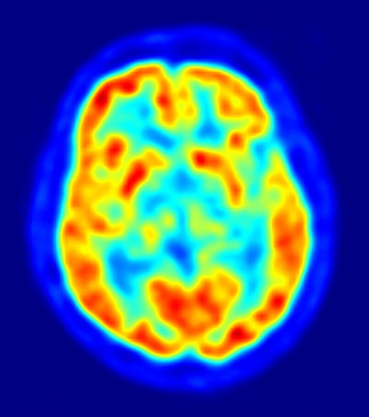
PET-image
An example of a PET scan of a brain. Credit to Jens Maus – http://jens-maus.de/


By Emma Yasinksi
BU News Service

In 2005, a serious car accident left a 23-year-old woman in a vegetative, unresponsive state.

For six months, she experienced sleep-wake cycles but was never able to communicate with her doctors and visitors. She couldn’t respond to simple commands such as “squeeze my hand,” or “look at this pen.”

Adrian Owen, Canada Excellence Research Chair in Cognitive Neuroscience at Western University, Canada wondered what was actually happening in her brain.

He slid her inside an fMRI, a machine that measures changes in the brain’s blood flow over time. Not knowing whether she could hear him, he told her to imagine she was playing tennis. He was excited to see that in response to his request, blood flow in her brain increased in the areas she would need to use to imagine herself playing the sport. While the blood flow in his patient’s brain was nowhere near that of a healthy individual, he felt that the increase was enough to indicate that she had heard him and was responding.

Over the past ten years, Owen’s “tennis test” has been replicated many times. Researchers have come to consider it a benchmark for studying vegetative patients, but it has never been applied as diagnostic tool. Now researchers are testing other imaging techniques that they hope will suggest which vegetative patients are most likely to awaken.

For now, clinicians are still using a scale developed in the 1970s to judge patients’ awareness based on their ability to respond to commands. The doctor will ask the patient to squeeze a hand, or look around the room. Then doctors use the responses to measure a patient’s diagnosis and likelihood of waking up on a scale which ranges from brain dead to conscious. Brain imaging studies using the tennis test have made it clear that doctors using behavioral methods are missing a group of patients who may be conscious but can’t respond to physical commands.

Up to 20% of patients who are diagnosed as vegetative using this scale actually pass the “tennis test,” according to Srivas Chennu, PhD, Senior Research Associate at Cambridge University, UK. This suggests that there is more going on in these patients’ brains than doctors might think.

Researchers are exploring two main imaging techniques that measure brain activity in vegetative patients. The first is the PET scan, which measures glucose in the brain. Johan Stender, graduate student at the University of Copenhagen, and his team PET-scanned the brains of 126 patients who were vegetative or minimally conscious (inconsistently able to respond to commands,) without asking the patients to imagine anything. They followed the patients’ progress for a year, to see who regained consciousness. The team compared the results of the PET scans to the results of the fMRI tennis test on the same patients. Those patients who showed activity in the PET scan also passed the tennis test, and were the most likely to wake up within a year. According to the study, published in The Lancet in 2014, The PET scan correctly predicted who would wake up three out of four times. The authors recommended using the PET scans to complement behavioral tests when diagnosing vegetative patients.

Another group, led by Chennu, aimed to find the most convenient and cost-effective way to measure brain activity in vegetative patients. They used eletro encephylograms (EEG’s) – administered at the bedside with a cap and some electrodes – to measure the electrical activity in patients’ brains. The EEG showed that 4 of 13 vegetative patients showed much more electrical activity than the others. They found that these same four patients passed the fMRI tennis test, suggesting that the EEG could also distinguish this type of consciousness. They published the study in PLOS Computational Biology in October of 2014.

Although these results need to be confirmed in a larger study, it seems all three types of brain measurements may be able to help clarify diagnosis of vegetative patients and possibly predict who will wake up within the year.

In an effort to gain more information from the convenient EEG test, Chennu’s team used graph theory, a type of mathematics designed to measure systems and networks to measure the strength of the activity in the patients’ brains. With more research, these rankings may give even more detailed information about an individual patient’s diagnosis

“[Families] want to know what the diagnosis really is so that they can move on and deal with that. Doubt and uncertainty are always bad things.” Owen told Nature in 2012. As studies move forward and validate tools and help doctors understand their patients’ level of consciousness, the technology promises to help families cope with heart-wrenching diagnoses as well.

New Parkinsons Drug Shows Positive Results in Rat Study

photo credit: Birth Into Being via photopin cc

By Emma Yasinski
BU News Service

Robin Williams was open about his struggles with depression and addiction, but only his family knew that he was facing another diagnosis at the time of his death – Parkinson’s disease. The seven to ten million people suffering from Parkinson’s can attest that the diagnosis is a harrowing one, foreshadowing an ever-changing cocktail of drugs, symptoms, and side effects for life. But, a new drug may have changed that- for rats at least.

Parkinson’s affects patients by killing neurons that make a neurotransmitter called dopamine in a small area, deep in the center of the brain, called the substantia nigra. Over time, patients struggle more and more to initiate movements. As the disease progresses, they may develop pain, depression, tremors, intestinal disorders, difficulty swallowing and confusion.

The most effective Parkinson’s drug on the market, levodopa, works by helping the brain replace the depleted neurotransmitter, but it cannot stop neurons from continuing to die. Doses must constantly be adjusted, which can lead to severe side effects. These side effects often need to be managed by separate drugs. Instead of struggling to initiate movement, the patient develops random, spontaneous movements. Levodopa unexpectedly stops working throughout the day, and the patient has to take more drugs with more side effects to keep it working. The patient and doctor must fight a decades-long, uphill battle to find a constantly changing combination of drugs and physical therapy.

Researchers at Emory University suspected that inflammation might be a key target for delaying the cell death in Parkinson’s disease. They developed a drug called XPro 1595 that prevents inflammation in the brain by binding to and blocking Tumor Necrosis Factor, a molecule that recruits inflammatory cells.

To test the drug, they used a well-known rat model of Parkinson’s disease in which the researcher induces the progressive, Parkinson-like cell death in the substantia nigra. Starting either 3 days or 14 days after creating the rat model, the researchers gave the rats XPro 1595 by intravenous injection in 3 day intervals.

After 35 days, the researchers evaluated the rats’ movements, levels of brain inflammation, and number of functional dopamine-producing neurons left in the substantia nigra. Rats that began dosage of the drug first (at 3 days) had the no noticeable movement disorders, minimal inflammation, and lost only 15% of their dopamine-producing neurons. The rats that began the regimen later were worse-off than the first group- with no movement problems, but some signs of inflammation and 44% of their dopamine-producing neurons gone. However, both groups were better off than the rats who received no drug at all. That group suffered motor symptoms, had the greatest signs of inflammation, and had lost 55% of their dopamine-producing neurons. Unlike current drugs, XPro 1595 was able to prevent cell death.

The human brain is capable of withstanding a lot of trauma, which makes it more difficult for doctors to diagnose Parkinson’s early. A patient rarely shows motor symptoms until 70% of these neurons are already dead. It would be too late for this drug to prevent the onset of symptoms.

That hasn’t stopped the Parkinson’s Disease Foundation from funding the next stage of research on XPro 1595, testing the drug in another well-known model of Parkinson’s in monkeys. Beth Vernaleo, Senior Manager of research programs at the Parkinson’s Foundation is confident that researchers are already beginning to identify tools for earlier diagnosis. It will still be four or five years before the drug enters clinical trials. “Our therapy is like a lady in waiting,” said Malu Tansey, Phd. a researcher involved in the study, “hoping that early identification of patients will give us an opportunity to test the efficacy of the drugs before people get motor systems.”